Contents
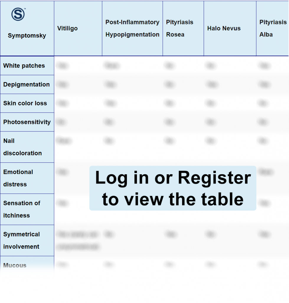
- 1 Vitiligo Differential Diagnosis Table:
- 2 How To Distinguish Vitiligo from Other Diseases
- 2.1 Distinguish Post-Inflammatory Hypopigmentation from Vitiligo – Diagnosis
- 2.2 Distinguish Pityriasis Rosea from Vitiligo – Diagnosis
- 2.3 Distinguish Halo Nevus from Vitiligo – Diagnosis
- 2.4 Distinguish Pityriasis Alba from Vitiligo – Diagnosis
- 2.5 Distinguish Tinea Versicolor from Vitiligo – Diagnosis
- 3 Important Red Flags in Vitiligo
Vitiligo Differential Diagnosis Table:

Vitiligo is an autoimmune disorder that causes the skin to lose its color; it becomes lighter than the skin tone or may turn white. These areas on the skin are called macules or patches.
Vitiligo occurs when cells called melanocytes, responsible for giving the skin its tone, get destroyed by the immune system.
The macules or the patches can affect any area on the body, including your hair, making the hair white.
The extent of symptoms of vitiligo varies differently from one person to another. In some people, it can affect small areas of the body, and for others, it can affect large areas, and there’s no way to predict the extent of the disease.
How To Distinguish Vitiligo from Other Diseases
Distinguish Post-Inflammatory Hypopigmentation from Vitiligo – Diagnosis
Post-inflammatory hypopigmentation is a condition where your skin becomes lighter and depigmented after inflammation in the skin from a disease or sun exposure. It usually happens in people with a darker skin tone. The depigmented parts are not regular or smooth like vitiligo, which is rather smooth.
- Diagnosis of this condition depends mainly on physical examination by the doctor and knowing the patient’s history.
- In some cases, a skin biopsy may be required to rule out other conditions or diseases such as vitiligo.
Distinguish Pityriasis Rosea from Vitiligo – Diagnosis
Pityriasis rosea is a self-limited rash; it appears on the skin as an oval red raised patch known as the herald patch. The exact cause of this rash is unknown but may be from viral infections, drugs, or vaccination.
- Diagnosis of pityriasis rosea depends mainly on physical examination and rash appearance because of the unique herald patch.
- If the doctor is not sure of the diagnosis, scraping of the skin or skin biopsy may be taken for histopathology to check for subacute dermatitis.
Distinguish Halo Nevus from Vitiligo – Diagnosis
Halo nevus is known as a melanocytic nevus; it’s a mole with hypopigmented edges and infiltrated with T-lymphocytes. Its exact cause is not known, but it may have an autoimmune origin. These moles can resemble melanoma, which makes definitive diagnosis important.
- If halo nevus is suspected, a full skin examination with a skin biopsy is needed to rule out or diagnose melanoma.
“Diagnosis of halo nevus can be confirmed by physical appearance, but due to the probability of melanoma, other invasive tests are required.”
Distinguish Pityriasis Alba from Vitiligo – Diagnosis
Pityriasis alba is a skin condition characterized by hypopigmented lesions with fine scales. These lesions may appear as red or pink patches at first but slowly fade away into hypopigmented lesions that can last several months and in some cases may last years. It’s a minor form of atopic dermatitis and often occurs in children and teenagers.
- Wood lamp examination can help identify pityriasis alba as it doesn’t lighten up or produce any fluorescence.
- A skin biopsy can help in diagnosis, but it’s rarely required.
Distinguish Tinea Versicolor from Vitiligo – Diagnosis
Tinea versicolor is a fungal infection that causes patches on the skin that can either be darker or lighter than your skin color. These lesions can be itchy but it’s completely harmless and not contagious, and full recovery is expected after treatment.
- In most cases, the doctor will scrape a part of the skin and diagnose it under a microscope to look for yeast cells.
- Wood lamp examination to see the skin under UV lights, tinea versicolor will appear yellow-green in color.
- An examination using KOH preparation can also be conducted, where the doctor will place a piece of infected lesion on a microscope containing 20% KOH.
Important Red Flags in Vitiligo
After the diagnosis of vitiligo, it’s important for the doctors to rule out any other autoimmune condition since the disease itself can be associated with other autoimmune conditions.
Vitiligo can affect ears, since it has melanocytes which can lead to hearing problems and sometimes hearing loss, up to 38% of patients can have auditory system involvement due to vitiligo.
Vision problems can also occur in vitiligo in areas of the eyes with melanocytes like retinal pigment epithelium; this can cause a change in vision and sometimes eye inflammation and redness can occur. Doctors usually recommend routine eye examination for vitiligo patients.
