Contents
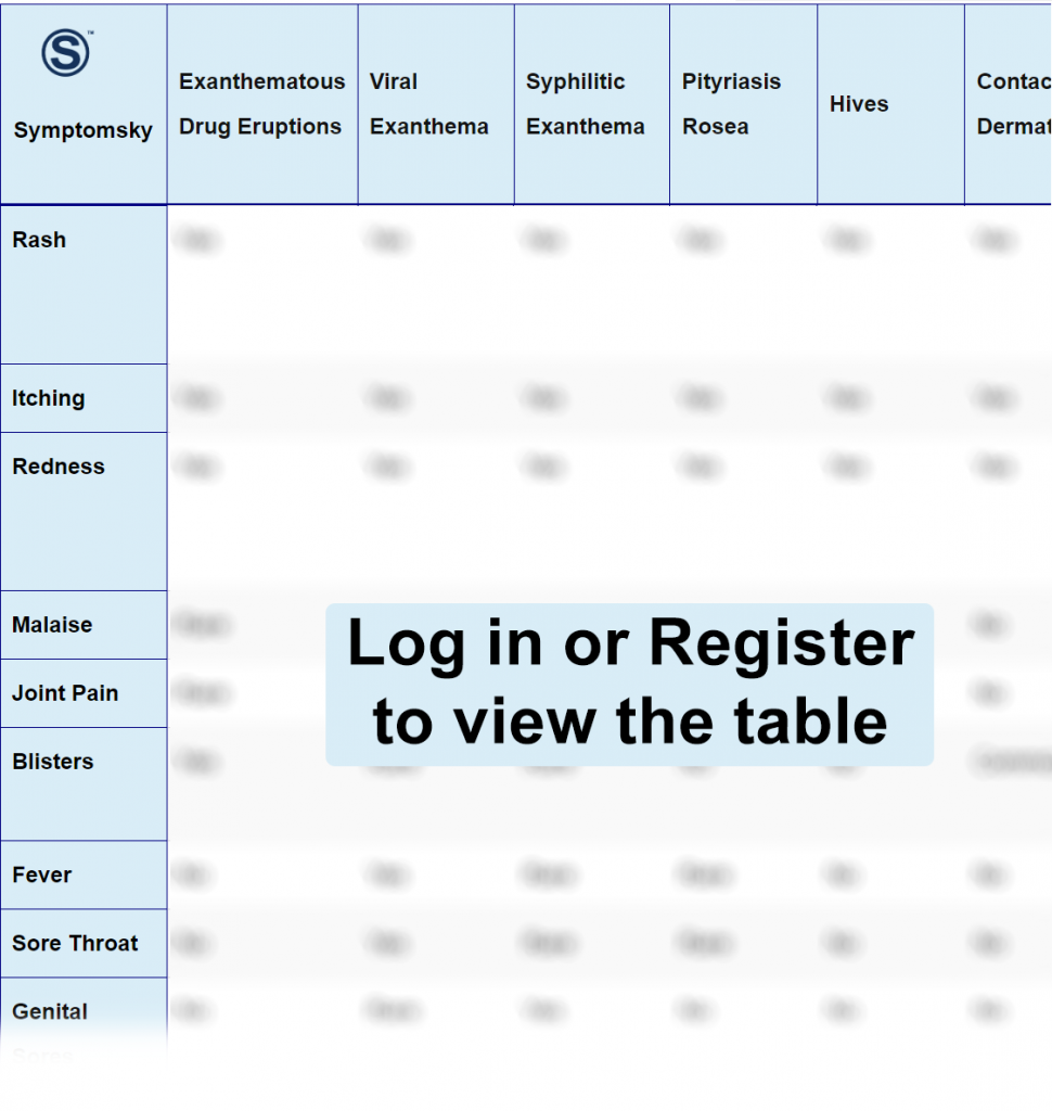
- 1 Exanthematous Drug Eruption Differential Diagnosis Table:
- 2 How to Distinguish Exanthematous Drug Eruptions from Other Diseases
- 2.1 Distinguish Viral Exanthem from Exanthematous Drug Eruptions – Diagnosis
- 2.2 Distinguish Syphilitic Exanthema from Exanthematous Drug Eruptions – Diagnosis
- 2.3 Distinguish Pityriasis Rosea from Exanthematous Drug Eruptions – Diagnosis
- 2.4 Distinguish Hives from Exanthematous Drug Eruptions – Diagnosis
- 2.5 DistinguishContact Dermatitis from Exanthematous Drug Eruptions – Diagnosis
- 2.6 Distinguish Purpura from Exanthematous Drug Eruptions – Diagnosis
- 2.7 Distinguish Scalded Skin Syndrome from Exanthematous Drug Eruptions – Diagnosis
- 2.8 Distinguish Photoallergic Dermatitis from Exanthematous Drug Eruptions – Diagnosis
- 3 Common Red Flags with Exanthematous Drug Eruptions
Exanthematous Drug Eruption Differential Diagnosis Table:

It is a toxicoderma in which well-defined round or oval-shaped erythematous plaques appear; they may present central blisters.
Exanthematous pharmacological reactions are adverse effects of hypersensitivity to a drug. The most frequent location is on the hands, feet, and genital area, when it resolves or leaves residual hyperpigmentation.
The lesions can be bright red macules or papules that converge to form large macules. They are always distributed over the trunk and extremities, but they can present greater confluences in fold areas.
How to Distinguish Exanthematous Drug Eruptions from Other Diseases
Distinguish Viral Exanthem from Exanthematous Drug Eruptions – Diagnosis
Viral Exanthem is a skin rash caused by a virus.
- Viral exanthem lesions are pink or red spikes that change color during pressure, while exanthematous drug eruptions lesions can be bright red macules or papules that converge to form large macules.
- Viral exanthem lesions appear over the trunk and neck and then spread to the arms, legs, and face, while exanthematous drug eruptions lesions appear on the site where the drug was applied or cause lesions over the body.
- Viral exanthem’s general picture consists of 2 or 3 days with high fever but good general condition; then, fever disappears, and a pink rash appears all over the chest, spreading into the face and trunk, unlike exanthematous drug eruptions where fever is rare.
- There may be pharyngeal inflammation, hyperemia, eyelid edema, and cervical adenopathy in viral exanthem, unlike exanthematous drug eruptions where fever is rare.
Distinguish Syphilitic Exanthema from Exanthematous Drug Eruptions – Diagnosis
Syphilis is an infectious disease that causes dermatological manifestations.
- Syphilitic exanthema lesions are round-shaped macules of pink-brown color and later on, well-defined dispersed papules, while exanthematous drug eruptions lesions can be bright red macules or papules that converge to form large macules.
- Syphilitic exanthema lesions appear 2-6 months after infection, unlike exanthematous drug eruptions lesions that appear at the moment or hours after the drug is administered.
Distinguish Pityriasis Rosea from Exanthematous Drug Eruptions – Diagnosis
Pityriasis rosea is a self-limiting inflammatory dermatosis.
- Pityriasis rosea’s most usual clinical presentation begins with oval salmon patches with a red necklace; later on, a rash appears accompanied by oval pink papules distributed in the form of a Christmas tree, while exanthematous drug eruptions lesions can be bright red macules or papules that converge to form large macules.
- Pityriasis rosea mostly affects adolescents and young adults, while exanthematous drug eruptions can affect people at any age.
- Pityriasis rosea has a seasonal predominance since it is more common during spring and autumn, while exanthematous drug eruptions can occur at any time.
- Pityriasis rosea patients may have general malaise, sore throat, and joint pain, while in exanthematous drug eruptions, it is very rare.
Distinguish Hives from Exanthematous Drug Eruptions – Diagnosis
Hives is an eruptive skin disease.
- Hives’ elementary lesion is the wheal, an erythematous lesion with elevation that has a clean center with a smooth surface, is not flaky, can be circular-shaped, circinate and presents confluence, while exanthematous drug eruptions lesions can be bright red macules or papules that converge to form large macules.
- Hives have a particular characteristic, and it is that the lesions disappear by themselves in less than 24 hours without leaving any residual lesion, unlike exanthematous drug eruptions that are only reversed using an antidote.
- Hives is an inflammatory entity; it can inflame different types of mucous membranes all over the body, causing systemic symptoms such as abdominal pain, vomiting, nausea, laryngeal edema, dysphonia, etc., while exanthematous drug eruptions rarely cause systemic symptoms.
DistinguishContact Dermatitis from Exanthematous Drug Eruptions – Diagnosis
Contact dermatitis is an inflammatory skin disease.
- Contact dermatitis appears after some external irritant or allergic agent touches the skin, while exanthematous drug eruptions require a drug into the system in order to produce a reaction.
- Contact dermatitis may also involve a slow evolution to scaling and thickening of the skin, while exanthematous drug eruptions lesions can be bright red macules or papules that converge to form large macules.
Distinguish Purpura from Exanthematous Drug Eruptions – Diagnosis
Purpura is a deadly blood disorder.
- Purpura is characterized by cutaneous hemorrhages due to a decrease in platelet count, while exanthematous drug eruptions are adverse effects of hypersensitivity to a drug.
- Purpura lesions are pinhead-shaped erythematous petechiae that do not change color by touch, are not palpable, and turn brownish as time passes, becoming yellowish-green, while exanthematous drug eruptions lesions can be bright red macules or papules that converge to form large macules.
- Petechiae are observed in the gingivae in Purpura, while in exanthematous drug eruptions, they are rare.
Distinguish Scalded Skin Syndrome from Exanthematous Drug Eruptions – Diagnosis
Scalded skin syndrome is a skin response to a toxin caused by a staphylococcal infection.
- Scalded skin syndrome mostly affects babies and young children, while exanthematous drug eruptions can affect people at any age.
- Scalded skin syndrome clinically looks like bullous impetigo, which presents scarce blisters and a scarlatiniform and generalized form, while exanthematous drug eruptions lesions can be bright red macules or papules that converge to form large macules.
- Scalded skin syndrome is caused by a bacterial agent, unlike exanthematous drug eruptions, which is a toxicoderma.
- Scalded skin syndrome main lesions are present in the navel, mouth, eyes, and anus, unlike exanthematous drug eruptions that occur mostly in the place where the drug is administered.
Distinguish Photoallergic Dermatitis from Exanthematous Drug Eruptions – Diagnosis
Photoallergic Dermatitis is a pathology in which skin cutaneous conditions occur whether due to exposure to solar or artificial radiation.
- Photoallergic Dermatitis is a type 4 immune reaction, unlike exanthematous drug eruptions, which is a toxicoderma.
- Photoallergic Dermatitis lesions are erythematous plaques that may present flaking with poorly delimited pruriginous erythema, while exanthematous drug eruptions lesions can be bright red macules or papules that converge to form large macules.
- Photoallergic Dermatitis lesions can persist even if the photosensitizing agent has been eliminated, while exanthematous drug eruptions lesions disappear when the drug that is causing it is removed.
Common Red Flags with Exanthematous Drug Eruptions
The drugs related to this type of reactions are: Penicillin, Sulfonamide, Benzodiazepines. These lesions can occur between 30 minutes and 8 hours after the medication. Patients can present: fever and chills.
Its more serious complication is the fixed bullous drug eruption consisting of blisters in multiple locations, similar to bullous impetigo.
