Contents
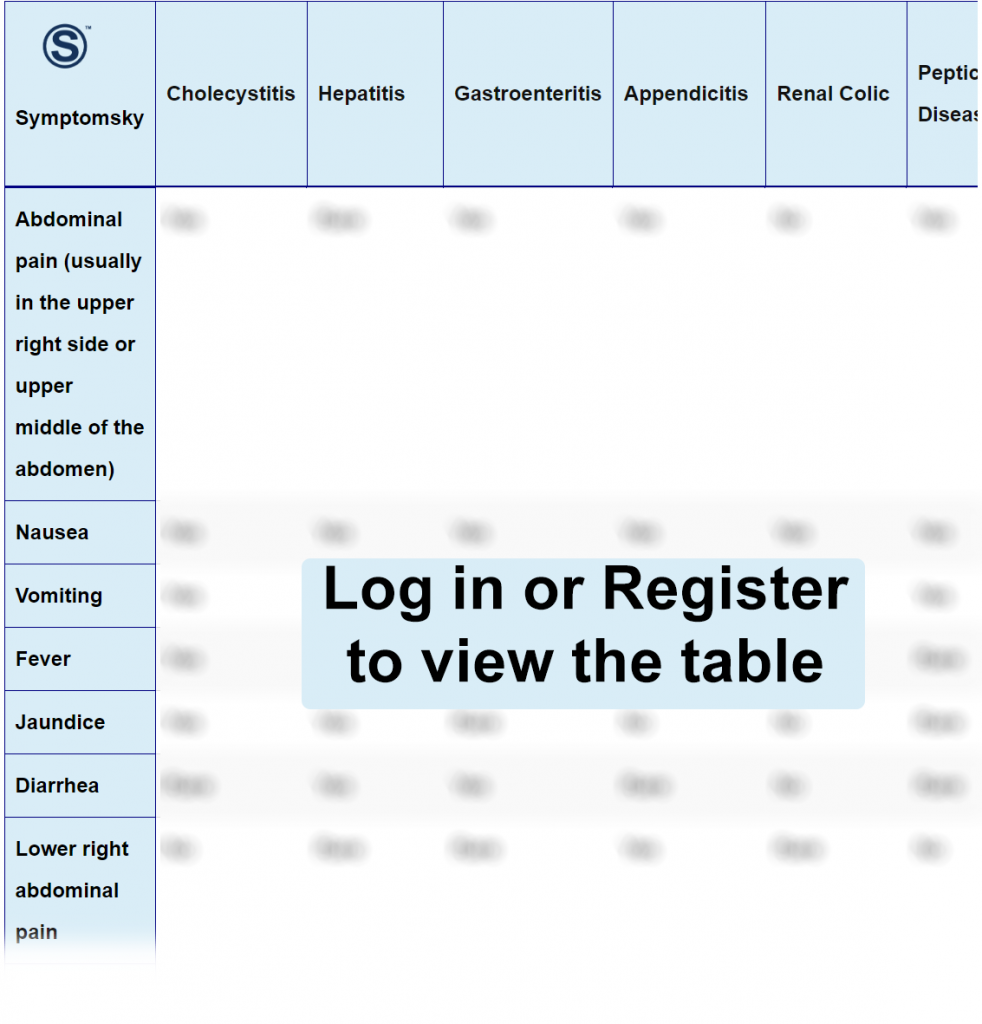
- 1 Cholecystitis Differential Diagnosis Table:
- 2 How To Distinguish Cholecystitis from Other Diseases
- 2.1 Distinguish Hepatitis from Cholecystitis – Diagnosis
- 2.2 Distinguish Gastroenteritis from Cholecystitis – Diagnosis
- 2.3 Distinguish Appendicitis from Cholecystitis – Diagnosis
- 2.4 Distinguish Renal Colic from Cholecystitis – Diagnosis
- 2.5 Distinguish Peptic Ulcer from Cholecystitis – Diagnosis
- 2.6 Distinguish Pancreatitis from Cholecystitis – Diagnosis
- 3 Important Red Flags in Cholecystitis
Cholecystitis Differential Diagnosis Table:

Cholecystitis is an inflammation of the gallbladder, which is responsible for the storage of bile to be secreted into the stomach for digestion. Cholecystitis happens when bile acids accumulate in the gallbladder due to a variety of reasons that can lead to blockage of bile ducts; gallstones, tumors, or even bacterial infection.
Cholecystitis is not often a life-threatening condition, but it can lead to severe pain. It can be treated either with medication or eventually surgery.
Cholecystitis can easily be diagnosed with imaging tests like CT, X-ray, and ultrasound. More specific tests for the gallbladder are like ERCP and PTC. Blood tests to measure total and direct bilirubin can also help to guide doctors for diagnosis.
How To Distinguish Cholecystitis from Other Diseases
Distinguish Hepatitis from Cholecystitis – Diagnosis
Hepatitis is an inflammation in the liver; it commonly occurs from viral infections like hepatitis A, B, C, D, or E, but it can also happen due to other causes like drugs and alcohol. Hepatitis is either an acute condition that may resolve on its own or with medication, or it can be a chronic condition that leads to serious hepatic injury, progressing to fibrosis or carcinoma. Cholecystitis itself can lead to hepatitis if left untreated for a long time.
- The first step usually for the diagnosis of hepatitis is liver function tests like ALT, AST, and alkaline phosphatase. (Even if not elevated, hepatitis is not excluded.)
- A virology panel of hepatitis A, B, and C is a must, as they are the most common causes of hepatitis.
- Ultrasound of the liver might be needed to determine any liver damage.
- Liver biopsy is also needed when other tests are negative and helps to determine the extent of liver damage.
Distinguish Gastroenteritis from Cholecystitis – Diagnosis
Gastroenteritis, also known as stomach flu, is a very common self-limited condition, usually arising from viral or bacterial infection. It causes stomach upset and severe vomiting and diarrhea, leading to dehydration, which is the most common complication of the disease. The condition itself goes away on its own.
- Stool test is the most common diagnostic test that can detect certain viral infections common for causing gastroenteritis.
- In most cases, the doctor can detect gastroenteritis just by physical examination and medical history.
Distinguish Appendicitis from Cholecystitis – Diagnosis
Appendicitis is an inflammation of the appendix, a very common disease that affects people at any age. It causes severe right lower abdominal pain, which is very characteristic of the disease. The pain is usually sudden, and most people will need immediate surgical intervention, but the procedure itself has a very low risk, and recovery and prognosis are very good.
- Physical examination can initially diagnose appendicitis; the doctor will apply pressure on the right abdomen, and pain will be relieved.
- CT scan is the confirmative test for appendicitis, which will visualize inflammation of the appendix.
- CBC is also maybe needed, which might show leukocytosis.
Distinguish Renal Colic from Cholecystitis – Diagnosis
Renal colic is the presence of kidney stones in the urinary tract, which causes severe lower abdominal pain. Although the most common cause of renal colic is a stone, sometimes spasms of the urinary tract can also lead to renal colic. This condition is very common, and usually, stones pass on their own with certain medications through the urethra, but if the stone is very large, surgical intervention may be needed.
- Imaging tests like ultrasound, CT, or X-ray can easily diagnose kidney stones and renal colic.
- Urinalysis is also needed to detect any urinary tract infection or substances like calcium that form stones.
Distinguish Peptic Ulcer from Cholecystitis – Diagnosis
Peptic ulcer is an inflammation in the lining of the mucous membrane that covers the stomach and protects it against acids. Sometimes, due to certain causes like H. pylori or overusing NSAID medication, that lining becomes inflamed, and it may progress to erosion and perforation, causing severe stomach pain and sometimes nausea and vomiting.
- Endoscopy is the most used test for the diagnosis of peptic ulcer. A tube with a camera in it will be inserted along the esophagus and stomach where any ulcer can be visualized, and tissue biopsy from the ulcer can be taken as well.
- A test for H. pylori, either stool antigen test or urea breath test, is routinely needed since most cases of peptic ulcer develop from H. pylori and to help guide treatment as well.
- Upper GI series, which is a series of X-rays with barium contrast, can sometimes be needed; it can give clear images of the ulcer.
Distinguish Pancreatitis from Cholecystitis – Diagnosis
Pancreatitis is an inflammation of the pancreas, that can be caused by a lot of reasons. One of the most common is cholecystitis itself, if progressed, it can cause inflammation in the pancreas. This can either be acute, which might be life-threatening if not treated fast, or can be chronic, which will progress over time, leading to permanent damage in the pancreas.
- Imaging tests like CT or X-ray can detect pancreatitis.
- More specific tests for pancreatitis like ERCP (can be used for management of pancreatitis as well) and MRCP are used too.
- Blood tests for amylase and lipase can help for guiding the initial diagnosis for pancreatitis.
Important Red Flags in Cholecystitis
Cholecystitis is often a treatable condition with good recovery and prognosis, but some red flags may present and reflect some serious complications.
Presenting with hemodynamic instability such as high-grade fever with tachycardia and low blood pressure may reflect gangrenous cholecystitis, in which gall bladder tissue has been necrotic, or may indicate perforation with peritonitis and sometimes sepsis as well.
The presence of jaundice as well, in which the patient will have yellow skin color and yellow eyeball, is very distinctive and reflects the prognosis of the disease and may indicate hepatic damage as well.
