
Contents
- 1 Bullous Pemphigoid Differential Diagnosis Table:
- 2 How to Distinguish Bullous Pemphigoid from Other Diseases
- 2.1 Distinguish Photo-Contact Dermatitis from Bullous Pemphigoid – Diagnosis
- 2.2 Distinguish Pemphigus Vulgaris from Bullous Pemphigoid – Diagnosis
- 2.3 Distinguish Erythema Multiforme from Bullous Pemphigoid – Diagnosis
- 2.4 Distinguish Systemic Lupus Erythematosus from Bullous Pemphigoid – Diagnosis
- 2.5 Distinguish Porphyria Cutanea Tarda from Bullous Pemphigoid – Diagnosis
- 2.6 Distinguish Bullous Fixed Drug Eruption from Bullous Pemphigoid – Diagnosis
- 2.7 Distinguish Dermatitis Herpetiform from Bullous Pemphigoid – Diagnosis
- 2.8 Distinguish Epidermolysis Bullosa from Bullous Pemphigoid – Diagnosis
- 2.9 Distinguish Linear IgA Dermatosis from Bullous Pemphigoid – Diagnosis
- 3 Treatment of Bullous Pemphigoid
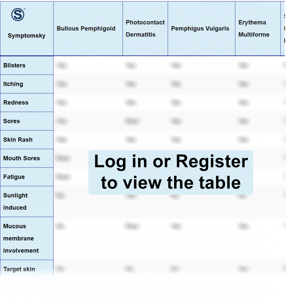
Bullous Pemphigoid Differential Diagnosis Table:
Bullous Pemphigoid is an autoimmune disorder that produces subepidermal blistering. Bullous pemphigoid is more common in elderly patients. It can be idiopathic or drug-induced with genetic predisposition. Some medications like diuretics, gliptins, TNF-alpha inhibitors, NSAID, and penicillamines are found to develop bullous.
The typical presentation of bullous pemphigoid includes a large, erythematous (red), itchy, and painful rash, which develops into bullae in weeks to months. These lesions are found commonly on the abdomen, trunk, flexor of arms, and medial thighs.
How to Distinguish Bullous Pemphigoid from Other Diseases
Distinguish Photo-Contact Dermatitis from Bullous Pemphigoid – Diagnosis
Photo-contact dermatitis develops from an interaction between sun exposure with a photo-reactive substance, while bullous pemphigoid occurs spontaneously.
- Photo-contact dermatitis develops from an interaction between sun exposure with a photo-reactive substance, while bullous pemphigoid occurs spontaneously.
“A photo patch test is done for differentiation of both diseases, as photo-contact dermatitis is a result of allergen and sun exposure.”
Distinguish Pemphigus Vulgaris from Bullous Pemphigoid – Diagnosis
Pemphigus vulgaris is an autoimmune blistering disorder.
- Pemphigus vulgaris can be found on both skin and mucosa, unlike bullous pemphigoid found on the skin only.
- Bullous pemphigoid can be differentiated from pemphigus vulgaris clinically by Nikolski sign where lateral pressure is applied to blisters, if splitting in the epidermal layer is positive Nikolski sign indicating pemphigus vulgaris, if the epidermal layers do not split negative Nikolski sign indicating bullous pemphigoid.
“Direct immunofluorescence can be used for differentiation between the two.”
Distinguish Erythema Multiforme from Bullous Pemphigoid – Diagnosis
Erythema multiforme is an acute, recurrent mucosal and cutaneous condition.
- Erythema multiforme can be found on mucosa and skin, unlike bullous pemphigoid, which is only found on the skin.
- Erythema multiforme appears as targetoid lesions, unlike bullous pemphigoid, which appears as blisters or bullae.
- Erythema multiforme lesions are not painful, unlike bullous pemphigoid, which is accompanied by pain.
“Immunofluorescence is used for differentiation between the two diseases.”
Distinguish Systemic Lupus Erythematosus from Bullous Pemphigoid – Diagnosis
Systemic Lupus Erythematosus is an autoimmune disease.
- Systemic Lupus erythematosus manifests systemically as fever, fatigue, and joint pain, which are absent in Bullous pemphigoid.
- Systemic Lupus erythematosus appears on the skin as malar and discoid rash activated by sun exposure.
- Lupus appears on mucosa as ulcers, but it does not develop bullae, unlike bullous pemphigoid, which appears only on the skin as bullae.
Distinguish Porphyria Cutanea Tarda from Bullous Pemphigoid – Diagnosis
Porphyria cutanea tarda is a dysfunction and alteration in heme biosynthesis.
- Porphyria cutanea tarda is followed by sun exposure, unlike bullous pemphigoid, which occurs spontaneously.
- Porphyria cutanea tarda leaves a scar after healing; bullous pemphigoid does not leave a scar.
- Porphyria cutanea tarda is differentiated by urine and plasma analysis, which shows the accumulation of porphyrin.
Distinguish Bullous Fixed Drug Eruption from Bullous Pemphigoid – Diagnosis
Bullous fixed drug eruption is an induced-drug condition.
- Bullous fixed drug eruption lesions start after drug ingestion within 24 hours, unlike bullous pemphigoid lesions, which take up to three months.
- Bullous fixed drug eruption and Bullous Pemphigoid can be differentiated by patch test (drug-allergen) and skin biopsy.
Distinguish Dermatitis Herpetiform from Bullous Pemphigoid – Diagnosis
Dermatitis Herpetiformis is a chronic autoimmune blistering disease.
- Dermatitis Herpetiformis starts due to gluten ingestion and is accompanied by gastrointestinal complications, which both do not occur in bullous pemphigoid.
“Dermatitis herpetiformis and Bullous pemphigoid can be differentiated through skin biopsy.”
Distinguish Epidermolysis Bullosa from Bullous Pemphigoid – Diagnosis
Epidermolysis Bullosa is a genetic inherited disorder causing keratinocytes mutation.
- Epidermolysis bullosa is present at birth or early childhood, unlike bullous pemphigoid, only found in elderly patients.
- Epidermolysis bullosa affects the skin and mucosa, while bullous pemphigoid only affects the skin.
- Epidermolysis bullosa causes defects in nails and teeth, which are both absent in bullous pemphigoid.
- Epidermolysis bullosa can also be differentiated through a magnetic microscope and immunofluorescence.
Distinguish Linear IgA Dermatosis from Bullous Pemphigoid – Diagnosis
Linear IgA dermatosis is a subepidermal condition causing vesicles and blister formation.
- Linear IgA Dermatosis is associated with vancomycin drug consumption, which does not occur in bullous pemphigoid.
- Linear IgA dermatosis has mucosal involvement, which is absent in bullous pemphigoid.
- Immunofluorescence is used for linear IgA dermatosis differentiation (IgA autoantibodies).
“Nikolski sign is used to differentiate between the two diseases; Linear IgA dermatosis has a positive Nikolski sign, but bullous pemphigoid has a negative Nikolski sign.”
Treatment of Bullous Pemphigoid
- Treatment depends on the disease severity and comorbidity. Corticosteroids are used as the first line of treatment.
- In more extensive severe cases, systemic prednisone is considered for disease limitation.
- Failing of corticosteroids for management, immunosuppressive therapy takes place.