Contents
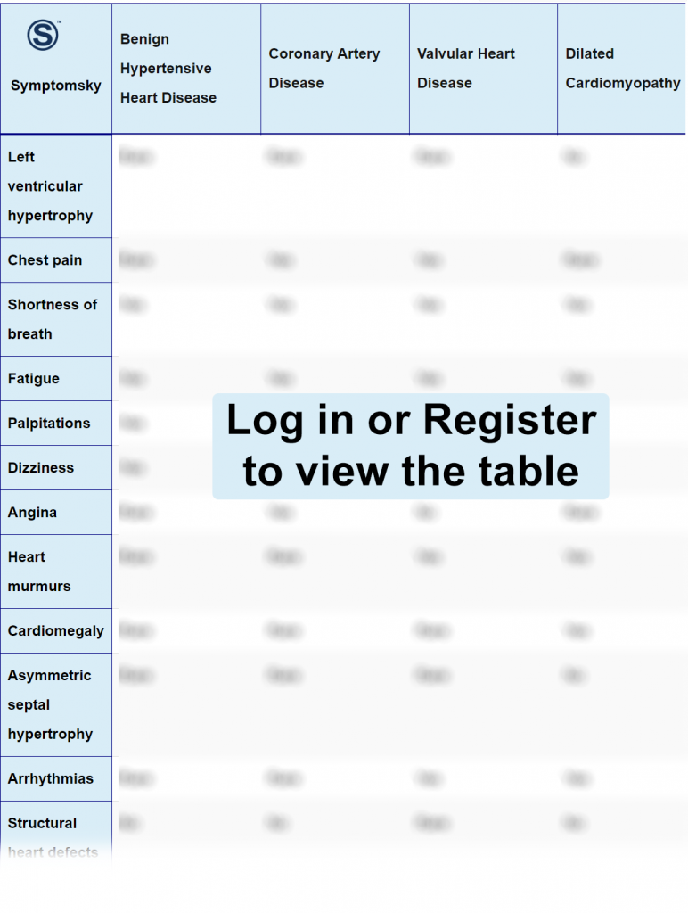
- 1 Benign Hypertensive Heart Disease Differential Diagnosis Table:
- 2 How to Distinguish Benign Hypertensive Disease from Other Conditions
- 2.1 Distinguish Coronary Artery Disease from Benign Hypertensive Disease – Diagnosis
- 2.2 Distinguish Valvular Heart Disease from Benign Hypertensive Disease – Diagnosis
- 2.3 Distinguish Dilated Cardiomyopathy from Benign Hypertensive Disease – Diagnosis
- 2.4 Distinguish Hypertrophic Cardiomyopathy from Benign Hypertensive Disease – Diagnosis
- 2.5 Distinguish Arrhythmogenic Right Ventricular Cardiomyopathy from Benign Hypertensive Disease – Diagnosis
- 2.6 Distinguish Congenital Heart Disease from Benign Hypertensive Disease – Diagnosis
- 3 Important Red Flags in Benign Hypertensive Heart Disease
Benign Hypertensive Heart Disease Differential Diagnosis Table:

Benign hypertensive heart disease is a chronic condition when the blood pressure is higher than 120/80 mmHg for a long period of time this can lead to heart problems over time, the exact etiology of hypertension is not known it can either be hereditary or from environmental factors such as smoking, obesity and increased salt intake.
Hypertension doesn’t usually cause symptoms that’s why it’s called the silent killer, most people found out they have hypertension during routine checkup or when blood pressure is so high that it causes headache and migraine.
Diagnosis of hypertension depends on measurement of blood pressure in more than one sitting and it’s higher than 120/80 mmHg. other tests like echo, ECG, electrolyte serum tests and kidney function is although needed to rule out any complications from disease.
How to Distinguish Benign Hypertensive Disease from Other Conditions
Distinguish Coronary Artery Disease from Benign Hypertensive Disease – Diagnosis
Coronary artery disease is a condition where coronary arteries become narrow leading to decreased blood flow to the heart, this can happen due to a variety of reasons but the most common is atherosclerotic plaque. Coronary artery disease has several types ranging from mild forms like stable angina to life-threatening conditions like myocardial infarction. Uncontrolled blood pressure for a long time itself can cause coronary artery disease.
- Echo can help in detecting wall motion abnormalities and abnormal blood flow to the heart.
- Coronary angiography helps in identifying narrowing of coronary arteries and assesses blood flow as well.
- ECG can help detect different types of coronary artery disease according to if T waves show abnormalities.
- Stress test can be used along with other tests to help more in diagnosis and accuracy of other diagnostic tools.
Distinguish Valvular Heart Disease from Benign Hypertensive Disease – Diagnosis
Valvular heart disease is when one or more valves in the heart become dysfunctional, either due to stenosis, prolapse, or regurgitation. This can lead to an enlarged heart or heart failure. This condition can either be managed medically or sometimes, if the case is severe, it might need surgical intervention.
- Echo is the main diagnostic tool for valvular heart disease and it can help in assessing the severity of the condition.
- ECG can also be used in the diagnosis of valvular heart disease, but early in the disease, it might not show any abnormalities.
- Imaging tests such as chest X-ray and heart MRI can show any problems with the heart such as an enlarged heart or structural problems in the valve.
Distinguish Dilated Cardiomyopathy from Benign Hypertensive Disease – Diagnosis
Dilated cardiomyopathy is a condition in which the wall of the heart becomes thin and stretched or dilated, affecting the heart’s ability to contract. Usually, it starts in one chamber of the heart and progresses slowly to other chambers. Medication may help in improving symptoms and slowing the progression of the disease.
- Echo is used for diagnosis, it’s the main test and can show abnormalities if the heart is enlarged.
- Chest X-ray can help visualize the heart and detect cardiomyopathy.
- Blood tests may be needed to detect the presence of infection since it can cause cardiomyopathy.
- ECG is routinely used to check for heart arrhythmia and monitor the prognosis of the disease.
Distinguish Hypertrophic Cardiomyopathy from Benign Hypertensive Disease – Diagnosis
Hypertrophic cardiomyopathy is a condition where the wall of the heart becomes thickened and stiff, making it very hard for the heart to contract and pump blood. Over time, this can lead to heart failure and arrhythmia.
- Echo is used for the diagnosis of hypertrophic cardiomyopathy, it can visualize an enlarged heart and any problems in the septum as well.
- Heart MRI and CT chest give detailed images of the heart.
- ECG is used to monitor heart rates and any signs of arrhythmia that may arise from the condition. Continuous monitoring and follow-up of heart rate are recommended.
Distinguish Arrhythmogenic Right Ventricular Cardiomyopathy from Benign Hypertensive Disease – Diagnosis
Arrhythmogenic right ventricular cardiomyopathy (ACM) is a genetic condition that causes defects in the heart, replacing normal tissue of the heart with fatty fibrous tissue. This disrupts the normal rhythm of the heart, leading to heart arrhythmia. This condition may lead to sudden death.
Diagnosis of ACM is usually hard, and there’s no specific test that can confirm the diagnosis.
- There are criteria to help doctors in diagnosis; the diagnosis is based on findings of 2 major criteria or 1 major and 2 minor criteria or 4 minor criteria.
- Genetic testing due to mutation of genes that are responsible for the disease can be positive in around 50% of patients.
- Angiography is a gold standard test for the diagnosis of ACM with specificity 90%.
- ECG can be used, but it has low specificity and sensitivity.
- Echo can visualize some abnormalities in the heart and complications from the disease.
Distinguish Congenital Heart Disease from Benign Hypertensive Disease – Diagnosis
Congenital heart disease is a structural heart disease that presents since birth and continues throughout life. This affects the quality of life and survival rate, but current treatment and surgery have improved prognosis.
- Heart MRI and CT scan can be used to give clear images of the heart and show any structural defects.
- Transesophageal echocardiogram gives detailed images of the heart and helps if normal imaging tests didn’t show any problems.
- ECG and echo are used for monitoring and follow-up of the condition and detect any complications that may arise like arrhythmia or cardiomegaly.
Important Red Flags in Benign Hypertensive Heart Disease
Hypertension is a chronic condition that requires continuous monitoring and follow-up. Hypertensive patients need lifelong medication for control of blood pressure to prevent complications and organ damage.
Common red flags in benign hypertensive heart disease include shortness of breath, headaches, dizziness, and fatigue. This may indicate that blood pressure is currently high and there’s a risk of stroke or angina, it’s an alarming sign for immediate checking of blood pressure.
Blurred vision is another red flag that may suggest hypertensive retinopathy, where the eye gets affected and may lead to permanent damage.
Hematuria and swelling of lower limbs especially may indicate that the kidney has been affected. It’s important in benign hypertension to control disease and prevent transformation to malignant hypertension (Hypertension with end-organ damage).
